


Ads by Google


color doppler CE certificate Adaptive sound speed optimization Support 2Dimageing multiply measurement
Detailed specification of 300 Full Digital Color Doppler
Focusing | Electronic focusing + acoustic lens focusing Transmission focus: single focus, double focus, 3focus and 4focus Continuously dynamic focusing mode is used for front end reception |
B-type display | Image amplification (real-time or freeze status), max. 4times |
Image vertical / horizontal overturn | |
Display depth is continuously adjustable | |
M-type display | Scanning speed: 4- stage |
Digital beam formation | Image whole range continuous dynamic focusing Image whole range dynamic aperture Image whole range dynamic trace change Image whole range receive delay weight summation Support semi-moment scanning, and support ± 10 ° linear reception deflection angle Multiple beam parallel processing technology |
Signal processing and Doppler | Provided with dynamic filtering and orthogonal demodulation Provided with total gain adjustment Gain adjustment: 8-segment TGC B-type, C-type, D-type total gain, respectively adjustable B/W image gain and color bloodflow gain, respectively adjustable Doppler stereo output volume adjustment : 0-255 Doppler baseline adjustment : 6-stage Pulse repetition frequency can be respectively adjusted: CFM PWD Provided with D-linear speed adjustment |
Image processing function | Image realizes dynamic range conversion and logarithm compression Image realizes time filtering Image realizes spatial filtering Image realizes frame correlation Image realizes edge enhancement Image realizes grey scale convertion Provided with selection of B|M-type or M-type scanning speed Provided with high-density and high frame frequency selection Provided with image convex array scanning angle, linear array scanning depth control Provided with image optimized processing: 6-stage Image left/right, up/down overturn Color grey scale bar inversion selection B/W image resolution 1024 × 768 × 8 bit (256 grey scale) Color image resolution: 1024 × 768 8 bit × 8bit × 8 bit color coding |
Basic measurement Calculation function | B mode basic measurement: distance, angle, perimeter and area(ellipse method, trace method), volume, stenosis, column diagram, cross-section diagram |
M-mode basic measurement: heart-rate, time, distance, speed | |
Doppler measurement: time, heart rate, speed, acceleration | |
Obstetrics measurement Calculation function | Gestational sac(GS), biparietal diameter (BPD), cephalo-rump length(CRL), femur length (FL), Humerus length (HL), Transverse Abdominal Diameter (TAD), backbone length (LV), occipitofrontal diameter (OFD), Abdominal circumference (AC), head circumference (HC), estimation of parietal transparent length, gestational age and expected date of childbirth. |
Obstetrics report | Measure and calculate amniotic fluid index(AFI) Calculate the rate (BPD/OFD, FL/AC, FL/BPD, HC/AC) Estimate fetus weight Infer the gesattional weeks and expected date of childbirth according to LMP , BBT Fetal Biophysical Score Fetus growth curve |
Gynaecology measurement Calculation function | Measurement and calculation of uterus, left ovary, right ovary, left follicle, right follicle etc |
Andrology measurement Calculation function | Measurement and calculation of prostate, testicle etc Calculation of prostate peculiar antigen predicted value PPSA, and prostate peculiar antigen density PSAD |
Urology measurement Calculation function | Measurement and calculation of left kidney, right kidney, bladder, residual urine volume etc. |
Peripheral blood vessel measurement Calculation function | Measurement and calculation of area stenosis rate, tube diameter stenosis rate |
Small parts measurement Calculation function | Measurement and calculation of thyroid, breast , enclosed mass etc |
Cardiac measurement Calculation function | Cardiac measurement software packet – providing analysis and measurement method for heart rate, speed, left ventricle, aorta, mitral valve, ventricle (right/left) Area stenosis rate percentage (%Area Sten), tube stenosis rate percentage (%Diam Sten) Body surface area (BSA). |
Memory Function | Probe parameters memory Image memory Cine memory Measurement result memory Report memory |
Cine function | Auto playback Manual playback Playback speed selection Retrieval playback Forward/reverse playback frame by frame |
Reports | Obstetrics report Gynaecology report Uology report Andrology report Cardiac report (left ventricle, aorta, mitral valve, ventricule ) Add ultrasonic imgae in report Directory management File management |
Grey scale | 256 Grey scale |
Input/Output interface | Network interface USB interface Video interface Serial communication interface |
Probes | Probe socket: 2 PCS Probe frequency: 2.5MHz~10.0MHz, 8-stage multi-frequency |
Probe class | Large convex array: frequency ranging between 2.0MHz and 10.0MHz, scan angle is 70 degrees. micro- convex array: frequency range of 2.0MHz to 10.0MHz, scanning angels ranging between 72 and 110 degees. high-frequency linear array: frequency ranging from 2.0MHz to 10.0MHz, scanning width ranges between 32 and 40 millimeters. transvaginal probe: frequency range of 2.0MHz to 10.0MHz, canning angle ranging between 140 and 158 degrees. |
Software upgrading functions | Available |
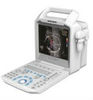
Liquid Crystal Display | SVGA line-by-line, flashless,15 inch high resolution LCD |
Power supply | Built-in switching power supply: input power ≤150VA Power supply adaption range: 220V ± 10%,50Hz ± 1Hz (AC |
Software Functions:
General measurement: distance, circumference, area, oval, trace, volume, HR, gestational week
Obstetric measurement soft: GS, CRL, BPD, AC, HC, FL, AFI, EDD, and calculating fetus weight, pregnancy week, expected delivery date, fetal physiological rating according to the previous measurements
Peripheral Ports:
VGA port
PAL-D video output
Specification:
Image mode | B, B/B, B+M, M, 4B, 8B |
Scanning mode | Convex and linear array |
Multi-frequency probe | 2.5~8.5 MHz |
Image magnification | ×1.0, ×1.2, ×1.5, ×2.0. |
Electronic focus | Acoustic lens focus, single focus and multiple focus |
Max display depth | 220mm |
Depth enhancement | Yes |
Local zoom | Yes |
Frame correlation function | Yes |
Adjustment total gain and dynamic range | Yes |
Gray Scale | 256 |
Display | 15'' LCD |
Gain Control | 6-segment STC segmentation control, full gain control |
Probe connectors | 1 (standard) |
Cine Loop | 256 frames |
Scanning frequency | 2.5~8.5 MHz (depends on probes) |
Image reverse | Left/right, up/down, positive/negative |
Comment | Name, Age, Sex,number of patient recorder, diagnostic date, 24 markers of body location, remarks in full screen |
Body marks | 18 |
Gray Scale Conversion | γ rectification |
Pseudo Color Function | 8 colors |
Power supply | AC 110V 60Hz,220V 50Hz |
Power consumption | ≤100VA |
Dimensions | 380x285x110mm (LxHxM) |
Net weight | approx. 6.8kg |
Standard Configuration:
Main unit
15'' LCD (abbr. Liquid Crystal Display)
256-frame cine loop
one probe connectors
3.5MHz electronic convex probe (2.5/5.0MHz)
Optional:
Electronic transvaginal probe (5.0/7.5MHz)
Electronic high-frequency linear probe (6.5/8.5MHz)
Thermal printer
compatible ultrasound scanner probe & color doppler machine